(To see other currencies, click on price)
MORE ABOUT THIS BOOK
Main description:
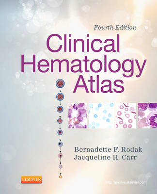
Ideal for identifying cells at the microscope, this atlas covers the basics of hematologic morphology, including examination of the peripheral blood smear, basic maturation of the blood cell lines, and discussions of a variety of clinical disorders. Over 400 photographs, schematic diagrams, and electron micrographs illustrate hematology from normal cell maturation to the development of various pathologies.
Contents:
Section 1: Introduction
1. Introduction to Peripheral Blood Smear Examination
Section 2: Hematopoiesis
2. Hematopoiesis
3. Erythroid Maturation
4. Megakaryocyte Maturation
5. Myeloid Maturation
6. Monocyte Maturation
7. Eosinophil Maturation
8. Basophil Maturation
9. Lymphoid Maturation
Section 3: Erythrocytes
10. Variations in Size and Color of Erythrocytes
11. Variations in Shape and Distribution of Erythrocytes
12. Inclusions in Erythrocytes
13. Diseases Affecting Erythrocytes
Section 4: Leukocytes
14. Nuclear and Cytoplasmic Changes in Leukocytes
15. Acute Myeloid Leukemia
16. Precursor Lymphoid Neoplasms
17. Myeloproliferative Neoplasms
18. Myelodysplastic Syndromes
19. Mature Lymphoproliferative Disorders
20. Morphologic Changes after Myeloid Hemopoietic Growth Factors
Section 5: Miscellaneous
21. Microorganisms
22. Miscellaneous Cells
23. Normal Newborn Peripheral Blood Morphology
Section 6: Body Fluids
24. Body Fluids
PRODUCT DETAILS
Publisher: Elsevier (Saunders)
Publication date: May, 2012
Pages: 296
Weight: 652g
Availability: Not available (reason unspecified)
Subcategories: Haematology, Midwifery
Publisher recommends


